
Interface Program Combining SPECT Images and MCNPX Code for Dose Calculation
Abstract
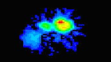
The paper describes a new interface program to convert Single Photon Emitted Computer Tomography (SPECT) images in Interfile format to Monte Carlo MCNPX code input file, and vice versa. The program was implemented in DELPHI 7.0, which opearates in a MS de Windows environment, and offers important tools that allow a flexible vizualization and manipulation of tomograhy slices. The material composition is computed in correspondence with the voxel geometry, given by SPECT images for an equivalent tissue. To validate the proposed interface,"S" factors were generated on a phantom of 81x81x81 voxels with a uniform activity distribution in the source voxel in Interfile format. The results were compared with those reported for the Y-90, as provided in the MIRD pamphlet No.17, which were calculated using the EGS4 Monte Carlo code. The comparison of results demonstrated good agreement (up to 2% difference) for the voxels next to the central source voxel. The program facilitates obtaining dosimetry calculations from SPECT images for users with little or no experience in the use of Monte Carlo codes, hence its convenience in routine clinical assessments. In order to demonstrate the program's applicability, images taken from a patient with hepatic metastases were used for the dosimetry calculations, which revealed satisfactory results.
This work is licensed under the Creative Commons Attribution-NonCommercial 4.0 International (CC BY-NC 4.0) license.